Evidence
We let the research speak for itself.
Simulation should earn its place in a curriculum. The modalities behind Deepscope have been evaluated in randomized controlled research and structured post-course assessment — the findings are summarised here, unedited.
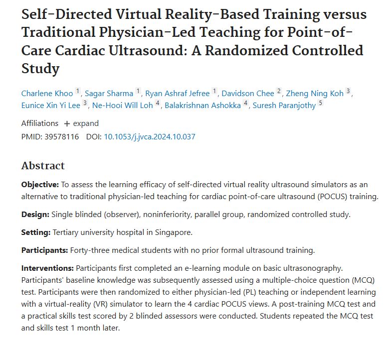
PMID 39578116 · DOI 10.1053/j.jvca.2024.10.037
Self-directed VR-based training versus traditional physician-led teaching for point-of-care cardiac ultrasound.
Key Finding
At 1 month post-training, self-directed VR training was noninferior to standard physician-led training… strong trends suggest enhanced retention of knowledge and procedural skills.
- Participants learned the four cardiac POCUS views either with a physician instructor or independently in VR.
- Skills were scored by two blinded assessors, with MCQ and practical testing repeated at one month.
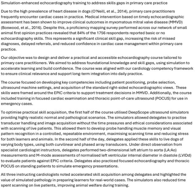
Simulation-enhanced echocardiography training in primary care practice.
Simulation-enhanced echocardiography training to address skills gaps in primary care practice.
Key Finding
Delegate confidence scores increased from a pre-course average of 2.4/5 to 4.5/5 post-course. All three instructing cardiologists noted accelerated skill acquisition among delegates.
- Simulators allowed transducer handling and image acquisition practice without the time pressures and ethical considerations of live scanning.
- Simulated pathology prepared learners for real-world cases while reducing time spent scanning live patients, improving welfare during training.
From the Field
What educators observe.
The quality of the cardiac anatomy is excellent. The ability to see how anatomical appearances change in response to transducer movements is particularly helpful for understanding relational anatomy and advanced cardiac views.
Irene Ma · Professor and John A. Buchanan Chair, University of Calgary
Participant feedback highlighted the practical format, clinical relevance, and the use of simulators as key strengths.
Course delegates · Simulation-enhanced echocardiography training
Our anatomical models are reconstructed from CT and MRI imaging, and imaging behaviour is validated against real transducer movement — so what learners see on screen changes faithfully as they scan.
We are committed to continued independent evaluation of Deepscope in medical and veterinary curricula. If your institution is interested in studying simulation-based ultrasound training, we would like to hear from you.
Evaluate it in your own programme.
Ask us about pilots, evaluation partnerships and the full study references.
Get in Touch