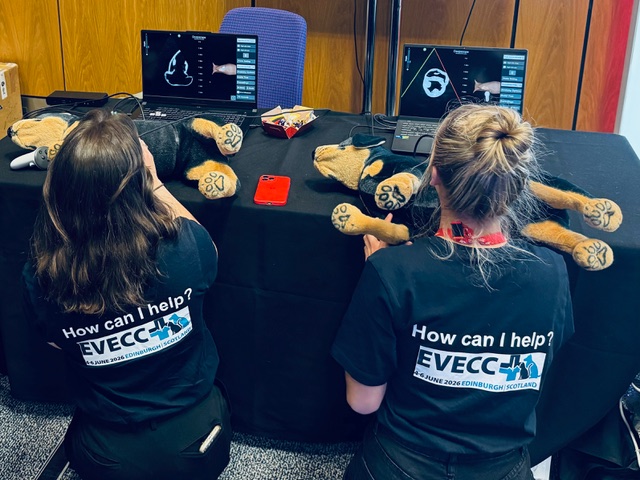
Mannequin Simulator
A tracked probe and physical mannequin provide genuine tactile feedback and psychomotor training. Instructors observe hand position while trainees visualise anatomy in real time.
ExploreMixed Reality Ultrasound Simulation
Deepscope turns any mannequin into a high-fidelity ultrasound simulator. CT and MRI derived anatomy, over one hundred anatomical structures, and a growing pathology library — portable enough to teach anywhere.
At a Glance
The Systems
Whether hands-on with a physical mannequin or fully immersive in virtual reality, every learner scans the same validated, standardised anatomy and pathology.
A tracked probe and physical mannequin provide genuine tactile feedback and psychomotor training. Instructors observe hand position while trainees visualise anatomy in real time.
Explore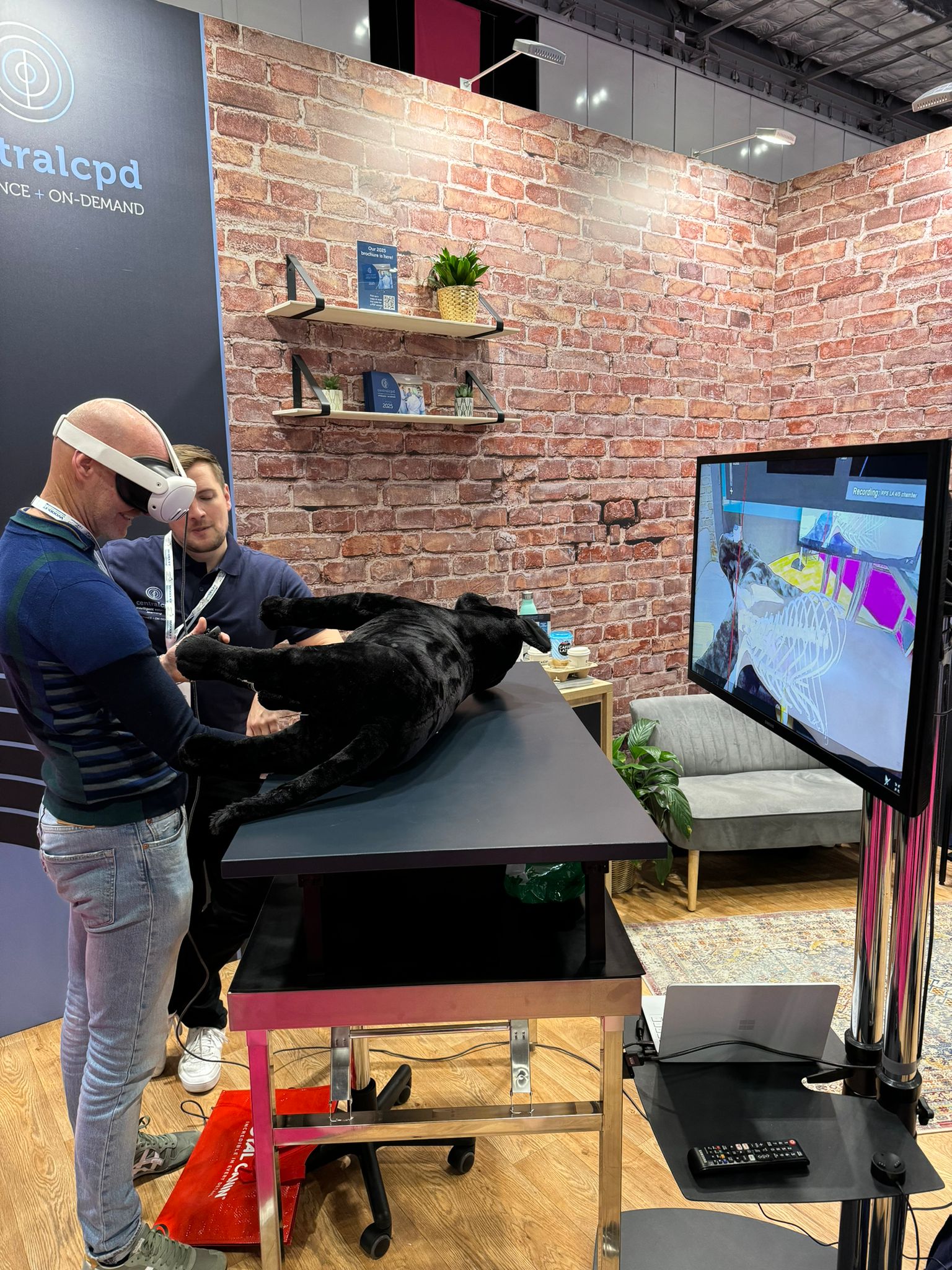
A fully immersive environment on off-the-shelf headsets. Virtual instructor guidance, on-screen calipers, and image capture support self-directed learning at cohort scale.
ExploreCapabilities
Position guidance for learning, position testing for assessment — objective measures of scanning technique.
Infarction, cardiomyopathies, valvular disease and more — every learner sees critical pathology before the clinic.
New pathologies are added regularly and included as free updates across all plans.
Headset, casting equipment and accessories in one case — simulation training, delivered anywhere.
Turn existing mannequins into high-fidelity simulators, including sliding on the body surface.
A medical simulation platform first, with dedicated veterinary anatomy and curricula as well.
Evidence
Deepscope modalities have been studied in randomized controlled research and post-course evaluation. We publish what we find.
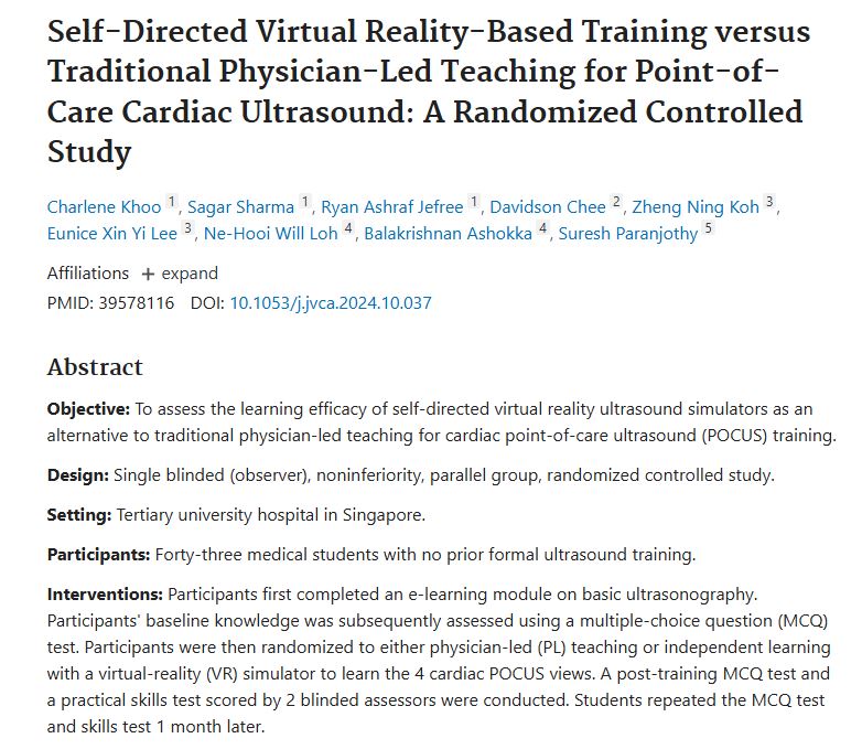
Randomized Controlled Study · Khoo et al., 2024
At one month post-training, self-directed VR training was noninferior to standard physician-led training, with strong trends suggesting enhanced retention of knowledge and procedural skills.
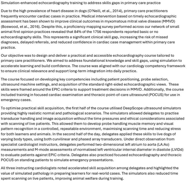
Simulation-Enhanced Echocardiography Training
Delegate confidence increased from 2.4/5 pre-course to 4.5/5 post-course. Instructors noted accelerated skill acquisition and highlighted the value of simulated pathology in preparing learners for real-world cases.
The quality of the cardiac anatomy is excellent. The ability to see how anatomical appearances change in response to transducer movements is particularly helpful for understanding relational anatomy and advanced cardiac views.
Irene Ma · Professor and John A. Buchanan Chair, University of Calgary
In Use At







Our mission is to help you teach more students, better. Tell us about your programme and we will be in touch.
Express Interest